The Zinc Linchpin Motif in the DNA Repair Glycosylase MUTYH: Identifying the Zn²⁺ Ligands and Roles in Damage Recognition and Repair

Figure: QM/MM energy-minimized structure of human MUTYH highlighting the four Cys residues interacting with Zn2+. Color coding as follows: C-terminal 8-oxoG recognition domain, pink; N-terminal adenine removal domain, green; interdomain connector, light blue; three previously identified Cys ligands (Cys318, Cys325 and Cys328 in human MUTYH), red; newly identified fourth Cys ligand (Cys230 in human MUTYH), purple; Cys residues coordinating the Fe-S cluster, orange, with the Fe-S cluster designated by orange (Fe) and yellow (S) spheres. DNA (from PDB ID: 5DPK) is in black with the azasugar transition state mimic (1N) in green, and 8-oxoguanine (8-oxoG) in pink, with elements depicted as hydrogen in grey, oxygen in red, nitrogen in blue, and phosphorous in orange.

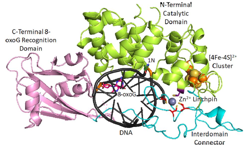
A recent publication from the David, Siegel and Lim (Academia Sinica, Taiwan) labs (Nuñez et al., JACS, 2018) provides insight into the coordination sphere and critical role of a Zn²⁺ metal binding site in the DNA repair glycosylase MUTYH. Genome database mining and sequence alignment of MUTYH orthologs, along with computational modeling, identified and supported Zn²⁺ ligation by four Cys residues. Three of the Cys residues lie in an interdomain connector region unique to mammalian MutY enzymes, while the 4th Cys is located in close proximity to the Fe-S cluster DNA binding domain. The functional consequences of reduced Zn²⁺ chelation on MUTYH-mediated DNA repair activity evaluated using a battery of in vitro and cell-based assays revealed the importance of Zn-coordination in recognition of the damaged DNA substrate. The critical nature of the “Zinc Linchpin Motif” suggests additional functions unique to higher organisms in damage signaling and crosstalk with other DNA repair pathways.

More information at https://pubs.acs.org/doi/10.1021/jacs.8b06923